
After injection of local anesthesia for root canal treatment, a healthy woman developed an extremely rare complication of traumatic myositis ossificans of the temporalis muscle insertion near the insertion of the right temporalis muscle.

After the dental procedure, the woman experienced a constant limitation in the width of her mouth opening. She eventually made a full recovery after undergoing a coronectomy, physical therapy, and regular jaw exercises, the authors write.
“This diagnosis is common in patients with treatment-resistant trismus following intraoral procedures,” wrote author Thorbjørn Østvik Pedersen, MD, Department of Oral and Maxillofacial Surgery, Haukeland University Hospital in Bergen, Norway.
Healthy woman aged 30
Before root canal treatment of the maxillary premolar, the woman was given local anesthesia in the area where the right temporalis muscle attaches. Immediately after the injection, the woman reported that she experienced severe pain and a persistent limitation in the width of her mouth opening after completion of treatment.
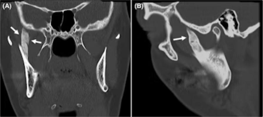
Four months after treatment, the woman developed an inability to open her mouth. By that time, her mouth opened only 8 mm. The patient could not move her jaw laterally. She underwent a computed tomography (CT) scan, which showed a bone spur approximately 3 cm long that extended from the right coronoid process of the jaw to the base of the skull.
(A) coronal and (B) sagittal CT scans show ossification of the insertion of the right temporalis muscle in a woman extending to the base of the skull (arrows).
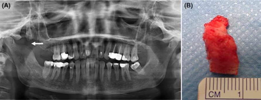
She underwent a coronectomy to partially remove the bone formation, and the sample was sent to the laboratory for analysis. The histological diagnosis of healthy bone tissue, combined with the results of computed tomography, led clinicians to the diagnosis of traumatic myositis ossificans.
Immediately after surgery, the patient had a mouth opening of 32 mm. After one month, the maximum mouth opening had decreased to 14 mm, although no osseous abnormalities were detected on imaging.
According to the report, after physical therapy and regular jaw exercises, at a postoperative visit five months after surgery, the mouth opening width was 27 mm and acceptable function was observed. The authors wrote that at the five-year follow-up, the woman's mouth opened 40 mm and she chewed normally.
A panoramic x-ray (A) after surgical treatment shows partial resection of the lesion and coronoid process (arrow). (B) Clinical image of the removed lesion.
A highly unusual condition
Traumatic myositis ossificans is a rare disease in which bone forms deep inside a muscle. This is a very unusual condition in the head and neck area. The muscle of mastication is most commonly affected, and most reported cases occur after minor surgeries such as third molar extraction.
When this does occur, symptoms usually appear three to six weeks after the local muscle injury. Additionally, there is limited research on morbidity following a dental procedure. However, the authors report cases of traumatic myositis ossificans affecting the medial pterygoid muscle after mandibular nerve block.
“This patient developed ossification of the temporalis muscle insertion after administration of local anesthesia, an extremely rare complication,” Pedersen and colleagues wrote.