
Imaging helped extract a denture from the intestines of a mentally handicapped 52-year-old woman who unknowingly swallowed it.
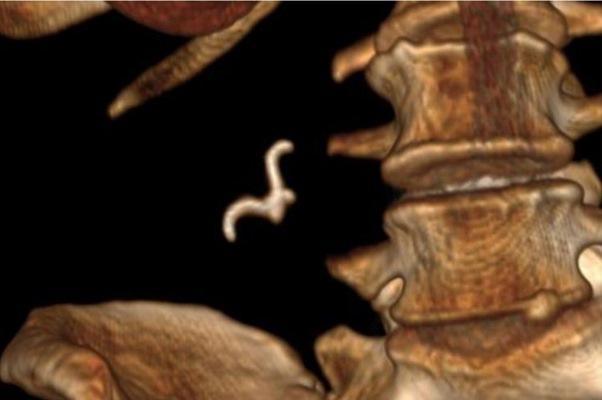
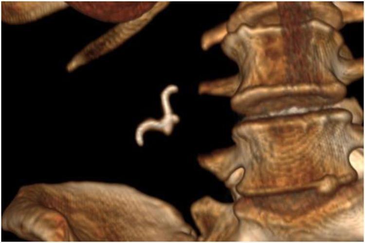
3D computed tomography (CT) reconstruction allowed clinicians to determine that she had swallowed a sharp, hooked object. This CT was the main tool in their surgical planning.
A team led by Dr. David Salvador of the Department of General Surgery at the Dr. José María Grande Hospital in Portalegre, Portugal, issued a statement that this case “raises awareness of the fact that diagnosis is often unpredictable, as even mentally capable patients may not notice or report swallowing foreign bodies, especially dentures.
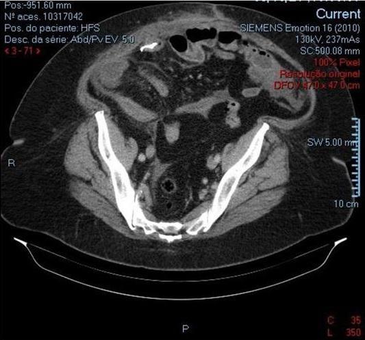
CT shows a solid metal object located in the small intestine in the right lateral quadrant with free fluid and localized fat thickening.
Foreign body ingestion is a common occurrence in the emergency department, but it is common in children, the elderly, or adults with cognitive delays or psychiatric disorders. To prevent serious complications, including bowel perforation or obstruction, clinicians should have careful discussions with patients and use imaging to determine the location of the foreign body and select the correct treatment plan.
In this case, a 52-year-old woman presented to the emergency department after 14 hours of gradual generalized abdominal pain and nausea. She had no fever, vomiting, or other symptoms. She had a history of diabetes, arterial hypertension and high cholesterol.
The examination revealed pain in the right iliac fossa of the woman, as well as peritonitis. The results of laboratory tests showed an increased number of leukocytes in the blood and the level of C-reactive protein. The chest x-ray showed no evidence of pneumatosis.
A 3D CT scan shows a hook-shaped object that turns out to be a denture.
A CT scan of the abdomen and pelvis showed an endoluminal metal object in the right lower quadrant, and doctors asked the patient if she had swallowed a fish bone or any foreign objects, including dentures. She didn't remember swallowing anything out of the ordinary.
In order to identify the object and the likelihood of intestinal perforation, they obtained a 3D computer reconstruction. The reconstruction showed that the foreign object had a sharp hook that could pierce the intestinal loop, so they decided to remove it surgically.
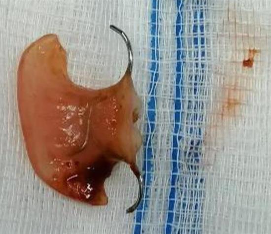
The surgeon immediately felt a hard object by touching the patient's bowel loops. Closer examination revealed an object embedded in the wall of the ileum with signs of impending perforation, approximately 20 cm from the ileocecal valve. The item, which was identified as part of a denture, was successfully retrieved.
A part of the denture was surgically removed from the patient.
Fortunately, the postoperative recovery of the woman passed without negative consequences. She was discharged six days after the operation.
Clinicians should ask patients with abdominal pain – regardless of mental health condition – about a possible foreign body and conduct imaging to rule out other conditions.
When diagnosing a patient with unexplained abdominal pain, ingestion of a foreign object should be present in the differential diagnosis, even in a mentally capable patient.