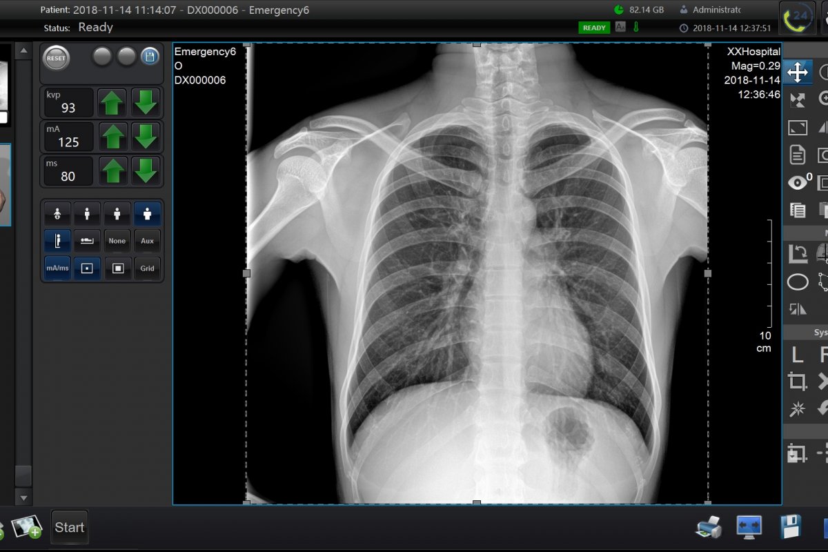
Historically, one of the first methods of recording X-ray radiation was X-ray-sensitive film. But its use had a number of significant drawbacks, because developing each image was a rather labor-intensive procedure.
It required the constant availability of special solutions with current expiration dates, operator skills for correct development and strict storage conditions (temperature, humidity, illumination, etc.). At the same time, the image existed only in a single copy, which caused great inconvenience during archiving and subsequent work with information.
A colossal breakthrough was the technology of computed radiography (CR). The radiation was recorded by a phosphor inside a special cassette, then the resulting image was read by a special scanner, the image was displayed in digital format on the doctor's workstation, and the phosphor was ready for the next image. The introduction of this technology freed operators from working with solutions, problems with storing and searching for information, reduced operating costs and opened up great opportunities in image processing.
The next evolutionary step was the emergence of digital flat panel detectors (DR). They allowed images to be obtained within a few seconds, did not require any actions to develop the image, had no parts subject to mechanical wear and significantly increased the contrast resolution of images. The thin, lightweight and durable design of the panels is convenient for work not only in the clinic, but also on the road.
And also DR panels:
– Faster than CR digitizers and film
The digital panel produces a picture faster than film or a CR system, since there are no stages of positioning the cassette in the digitizer or the film in the developing machine. The image automatically appears on the monitor.
– Highly detailed
The digital detector provides better image detail with a lower radiation dose. Thus, the use of DR panels reduces the radiation load on the patient and the doctor.
– Can work outside the clinic
A set in the form of a laptop, DR panel and portable X-ray machine is convenient to take on trips. If the detector is equipped with a Wi-Fi module and battery, then no wires are required during operation.
What are they?
Spoiler: there will be a lot of complex terms now.
Flat panel detectors for radiography are divided into two main types: based on CMOS technology and based on TFT technology. TFT detectors are divided into two subtypes: direct and indirect, and CMOS detectors are only indirect.
Thin-film transistors (TFT technology)
The operating principle of indirect TFT detectors:
- A scintillator based on cesium iodide or gadolinium oxysulfide converts X-ray photons into light radiation;
- A photodiode array, usually made of amorphous silicon, converts the light signal into an electrical signal;
- The electrical signal is recorded pixel by pixel by an array of thin-film transistors, processed and converted into an image.
The operating principle of direct-type TFT detectors:
- A scintillator based on amorphous selenium converts X-ray photons directly into an electrical signal;
- The electrical signal is recorded pixel by pixel by an array of thin-film transistors, processed and converted into an image.
In simple terms, direct conversion detectors do not involve the step of converting X-rays into light, or light into electrical energy. This avoids additional signal losses, making these detectors very sensitive to low radiation doses. The devices are common in areas where high spatial resolution is required at low radiation exposure, such as mammography.
CMOS-based detectors
An alternative to an indirect flat panel detector is CMOS or CMOS (complementary metal-oxide-semiconductor) technology, which is used in digital cameras, among other things.
In such detectors, the first stage of processing is similar to indirect detectors – X-ray radiation is converted into light using a scintillator. But then the following happens: using a bundle of optical fibers, a focusing lens or an electron-optical converter, the light is focused on a CMOS matrix, and then converted into an image by it.
Previously, a strong advantage of such devices over TFT detectors was the speed of reading information from the matrix. This determined their place in fluoroscopy devices.
Today, detectors based on TFT technology have a high speed of reading information, which also allows them to be used for fluoroscopy with a high frame rate. Thanks to this, a new class of devices has appeared – dynamic TFT detectors.
How to choose?
When choosing a flat panel detector for digital radiography, it is important to consider:
1. Length and width of the active surface
Standard sizes: 17×17″ or 14×17″. A larger active surface allows you to cover a larger area of the study. This helps in more accurate diagnostics and determining the nature of the pathology.
2. Detector pixel size
The parameter directly affects the quality of the resulting image: the smaller the size of one pixel, the higher the resolution of the device. Most modern panels have a pixel size in the range of 100-200 µm.
3. Availability of automatic response technology
The parameter is directly related to the ease of use and examination time: the detector switches to the image standby mode immediately after the start of the examination without an additional command from the operator. This is especially important when conducting examinations in several projections.
4. Software
Convenient software and the presence of auxiliary functions help the specialist in his practice.
5. Method of connection to the workstation: wired or wireless
Wired connection provides more reliable data transfer between the detector and the workstation: there is no risk of external interference or signal loss. It is relevant in stationary conditions, where there is no need for mobility.
Wireless connection has advantages in mobile conditions or when the detector needs to be moved frequently. It provides more flexible operation, ease of use and no wires.
6. Scintillator: CsI or GOS
The advantage of GOS or gadolinium oxysulfide is its lower cost. But the crystals of the substance have a complex structure, which leads to the effect of light scattering.
Cesium iodide CsI crystals have a columnar structure. This allows light to pass through, as if through fiber optic channels. Higher conversion efficiency provides higher image quality with less radiation exposure.
Where to buy?
Specialists from the East Medical Group company will help you buy a suitable flat panel detector.. Since 2006, we have been equipping state centers and private clinics with equipment for functional diagnostics and resuscitation.
Our catalog presents two models of the DR system PZ Medical: 1417 and 1717. The design of both uses TFT technology, CsI is used as a scintillator, which guarantees the best image quality.
The PZ Medical team consists of experts in the field of microelectronics and digital imaging. Their products have received CFDA, CE and FDA certifications. Each detector meets the highest quality standards.
You can view the equipment before purchasing in our showrooms in Moscow, St. Petersburg, Krasnodar and Novosibirsk.
To place an order or get advice, leave a request on the website, send a request to sale@e-medical.ru or call the toll-free number 8 (800) 5003085.
Advertisement: OOO East Medical Group
INN 7735178032, erid: 2SDnjbuKFtn
Important! Information provided for reference purposes. Ask a specialist about contraindications and side effects and do not self-medicate under any circumstances. At the first signs of illness, consult a doctor.
