
Due to the natural curvature of the spine and the high mobility of the cervical region, intervertebral hernias often occur in it. This is the destruction of the cartilaginous membrane of the disc, which separates one vertebra from another. In this case, the contents of the disk come out. Let's look at a real clinical case, why a hernia causes discomfort, how it is diagnosed and eliminated.
Signs of pathology
When the cartilaginous membrane of the disc is destroyed, its internal contents – the liquid core – comes out. It acts on the nerves in two ways:
- irritates chemically – by the same principle, the skin hurts when acid gets on it;
- presses mechanically – an impulse travels worse along a pinched nerve, just like current flows through wires.
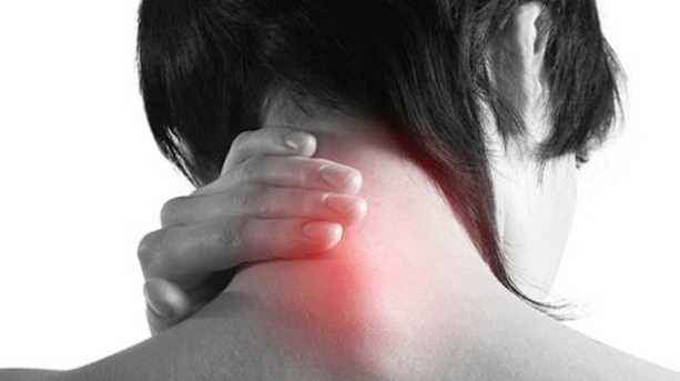
Due to nerve damage, characteristic symptoms occur, depending on the area of u200bu200bdamage.
Characteristic signs of damage to the cervical segment:
- back pain radiating to the leg or arm;
- numbness or weakness in the arm or leg;
- no ability to raise a straight outstretched leg while lying down.
Diagnostics
Changes are best seen with magnetic resonance imaging – MRI. They reflect the state of the disks themselves, as well as the level of compression of the nerve fibers, muscle swelling and the state of the paravertebral joints. The images of the patient showed a large lateral hernia on the right side at the level of 6-7 cervical vertebrae.
If the patient has metal or electronic prostheses, including pacemakers, they do not undergo an MRI. Such patients are prescribed computed tomography – CT. A 3D model of the spine is taken layer by layer on a computer and the changes are recorded. Sometimes a contrast agent is additionally injected to make the pathology better visible.
Treatment methods
Usually, therapy begins with conservative methods. This is suitable if the pain is not constant, and the sensitivity and function of the hands and feet are not impaired.
Methods of conservative therapy: NSAIDs) relieve pain and inflammation, and if they do not help, the doctor may offer strong prescription painkillers of the narcotic group under the supervision of specialists; fibers, or foraminal – to the place where the nerve root comes out;
Most patients are prescribed conservative therapy for 4-6 weeks, and only if it is ineffective, surgical methods are offered. So it was with the girl in question with a hernia in the cervical region – she was treated with medication for 3 weeks, but the pain was constant and too strong. When patients are so worried about pain, they are immediately prescribed surgery. At the Professor Sampiev Center they are operated on out of turn, the next day after the appeal.
The patient underwent a classic operation for a hernia of the cervical region using an anterior approach. A Swiss cage with two screws was installed in place of the destroyed disk. For the neck area, a zero-profile cage is chosen so that it does not come into contact with vessels, nerves, and organs adjacent to the front. With its help, the vertebrae were stabilized and the required height between the vertebrae was provided.
After the operation, the pain immediately disappeared, only a slight numbness of the arm remained. Over time, it will also go away if the woman undergoes rehabilitation to restore nerve conduction. Due to the fact that it was clamped for a long time, some of the cells died – it takes time for their regeneration. Numbness can persist for up to a year.
Three days after the operation, the patient was discharged home. Such short recovery times are possible thanks to high-tech equipment and modern techniques, as in the Center for Orthopedics of Professor Sampiev.
Advertising. Kra23XMzj