
Bioprinting and advances in materials are facilitating the creation of collagen structures needed for oral and maxillofacial surgery.
Despite the fact that modern methods of treating periodontal diseases offer enough solutions for restoring tooth tissues, they often cannot reproduce the original complex anatomical structure of the tissues. By providing precision tissue regeneration, 3D bioprinting has revolutionized this field. A team of scientists from China has published a review of currently available collagen-based 3D bioprinting methods and their applications for oral tissue regeneration. Namely, pulp and blood vessels, cartilage and periodontal tissue.
3D printing, pioneered by Charles Hull in 1986, uses digital models to create objects layer by layer using adhesive materials, providing design flexibility, reducing manufacturing costs and expanding functionality. In dentistry, 3D bioprinting aims to regenerate oral tissue and has changed the direction of dental surgery towards more precise and digitized approaches.
Collagen, a major component of the extracellular matrix, is often used in tissue engineering due to its structural and chemical similarity to the extracellular matrix of oral tissues. However, its rapid degradation limits its direct application in tissue scaffolds. To increase its effectiveness, collagen is usually combined with other materials. For example, to create biomimetic structures, researchers combined bioactive glass with modified collagen to improve the viability of human mesenchymal stem cells (MSCs).
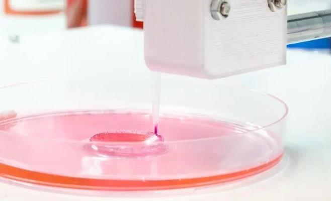
Collagen-based bioinks provide excellent printability across a variety of 3D printing methods. The suitability of collagen for printing depends on the composition of the bioink and the printing technology. 3D printed collagen scaffolds can provide an environment for cell growth, either by attracting stem cells or by directly housing the cells.
Bioprinting methods
Bioprinting technologies are broadly classified into inkjet, pressure-assisted bioprinting, and light-assisted bioprinting. Bioprinting, based on traditional inkjet printing, deposits ink droplets layer by layer. It enables precise placement of cells into 2D and 3D structures suitable for tissue engineering. However, when printing high-density cells, nozzle clogging can cause problems.
Extrusion bioprinting uses pneumatic or mechanical pressure to push the biofilm through a nozzle. The print is scalable and works well on highly viscous biofilms. However, cell viability can be affected by factors such as nozzle diameter.
First introduced to create 2D cell models, light-based bioprinting uses lasers to cure bioinks, providing minimal mechanical stress and allowing the creation of high-resolution structures. Techniques include laser-induced direct transfer (LIFT), stereolithography, and digital light processing. These methods allow the creation of scaffolds with high structural accuracy and good cell viability.
Practical application
Studies on 3D bioprinting of cartilage using collagen bioinks have shown that high-density collagen hydrogels, when heated, increase the geometric accuracy of the printed structures, improving structural precision and mechanical strength. Another similar study compared different cartilage-printing biofilms and found that alginate–agarose and alginate–collagen combinations provided superior compressive and tensile strength compared to alginate alone. In addition, alginate–collagen promoted better cell survival and effectively maintained the chondrocyte phenotype.
3D printing has also proven useful in recreating the nanofibrous structures of cartilage extracellular matrix. These scaffolds were found to support cell adhesion, proliferation and differentiation and demonstrate osteochondrosis regeneration potential. There have also been initiatives to use 3D printing for temporomandibular joint reconstruction. Gelatin-derived scaffolds have demonstrated potential for the differentiation of human bone marrow-derived MSCs into cartilage. Research has also highlighted the effectiveness of using both methods simultaneously, finding that such structures promote bone and cartilage growth.
Collagen Extraction
There are several methods for extracting collagen. A highly efficient and environmentally friendly extraction method is enzymatic extraction, which breaks down the covalent bonds in collagen under acidic conditions, preserving its physical and biochemical properties of collagen. Commonly used enzymes include pepsin, pancreatic protease, papain and ficin. Factors affecting extraction include temperature, pH, time and enzyme concentration. In addition, sophisticated extraction methods have been developed to achieve efficient and rapid extraction of collagen without compromising the structure.