
Assessing the bone mineral density (BMD) of the sides of the jaw, known as the mandibular ramus, using panoramic dental radiography can help identify patients at risk of developing osteoporosis.
The study authors write that panoramic dental X-rays may serve as an additional screening tool for osteoporosis.
“These results highlight the value of panoramic radiography as an additional method to identify individuals at risk for osteoporosis, especially when assessing BMD in areas such as the mandibular rami that are free of dental intervention and significant muscle forces,” write the authors, led by Dr. Alan Grupioni Lourenco, PhD, of the University of Ribeirão Preto School of Dentistry in São Paulo, Brazil.
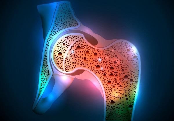
Osteoporosis, which commonly occurs in postmenopausal women, significantly reduces bone density and increases the risk of fractures. Cortical bone, which is the largest accumulation of calcium in the human skeleton, is primarily affected by diseases such as osteoporosis. Since cortical bone is visible on panoramic radiographs, this area of the mandibular canal can be useful in assessing BMD.
To characterize and compare changes in the cortical areas of the mandibular canal, panoramic radiographs of 52 postmenopausal women with normal osteopenia and osteoporotic syndrome over the age of 40 were analyzed. All women underwent osteoporosis risk assessment using dual-energy X-ray absorptiometry (DEXA).
Of these women, 26 had good radiographs, 19 had osteopenia, and 8 had osteoporosis. In this cross-sectional study, the intensity of black pixel on radiographs was used to measure BMD of the mandibular canal cortex.
Significant differences were found between groups, namely in the percentage of black pixels in the mandibular rami. For the good radiograph group, the average percentage of black pixels was 3.19% (±0.65). In the osteopenia and osteoporosis groups, the average percentages were 2.78% (=0.65) and 2.35% (=0.65) (p = 0.015), respectively.
However, the study had its limitations, including the small number of patients with osteoporosis in the study. The limited number of patients made it difficult to generalize the findings.
In the future, the authors plan to conduct a bivariate analysis of covariance based on pixel density by region to better confirm these results.
“We found statistically significant differences in mandibular ramus pixel intensity between patients classified as healthy and osteoporotic based on DXA results,” Lourenco et al. write.