
According to the report, while treating peri-implantitis, air was introduced into the skull of a 62-year-old woman, which is life-threatening. This happened when a woman was undergoing a submucosal debridement procedure to treat a peri-implant lesion using a special air polishing device.
Two seconds after turning on the device, the patient complained of severe discomfort in the face and head. Computed tomography (CT) revealed the presence of air in the woman's intracranial space, a phenomenon called pneumocephalus.
Since the air was probably contaminated with oral bacteria, the patient was in danger of developing meningitis. She was immediately admitted to the hospital and recovered after being treated with antibiotics.
“The present case of direct development of pneumocephalus as a result of a dental procedure is the first reported in the medical literature and should therefore be considered an extremely rare complication,” write the authors, led by Dr. Corinna Bruckmann from the Department of Conservative Dentistry and Periodontology, University Dental Clinic, Medical University of Vienna, Austria.
Very rare complication
In 2019, a 62-year-old woman with no noticeable health problems enrolled in a routine peri-implantitis treatment program at the Medical University of Vienna. She had had a sinus lift around teeth 26 and 27 five years earlier. Postoperative healing was uneventful and two implants were placed.
At the time of admission in 2019, the woman had a probing pocket 6-7 mm deep, bleeding during probing, and purulent discharge in the area of implants. Over the next few months, she underwent two courses of non-surgical peri-implant therapy using manual and ultrasonic instruments in addition to additional systemic and topical antibiotics.
The treatment was then interrupted due to the quarantine associated with the COVID-19 pandemic. When she resumed treatment in March 2021, her pocket depth was again 7mm on Implant 27. The clinic decided to remove the supragingival and subgingival biofilm using a supragingival air polisher with erythritol powder.
Following the manufacturer's recommendations, the device was positioned at a 60° angle at a distance of 3-5 mm towards the gingival sulcus. The device was set to 100% water flow and 40% power. During the procedure, a sterile disposable nozzle was used.
The device was activated when the tip was inserted mesially to a depth of 7 mm at implant 27. Two seconds later, the patient reported severe discomfort in the left side of her face and head, and treatment was stopped.
The patient was placed in an upright position. She was conscious and reported brief shortness of breath, pain behind her left eye, and difficulty swallowing. Blood pressure, heart rate, and pupillary response to light were normal. Extraoral examination and intraoral examination revealed nothing unusual.
Due to the strange sensations reported by the patient and unclear clinical signs, the patient underwent a CT scan, which showed the development of subcutaneous emphysema. The air spread from the front side in the caudal and cranial directions. Air was also found in the left carotid canal and inside the skull.
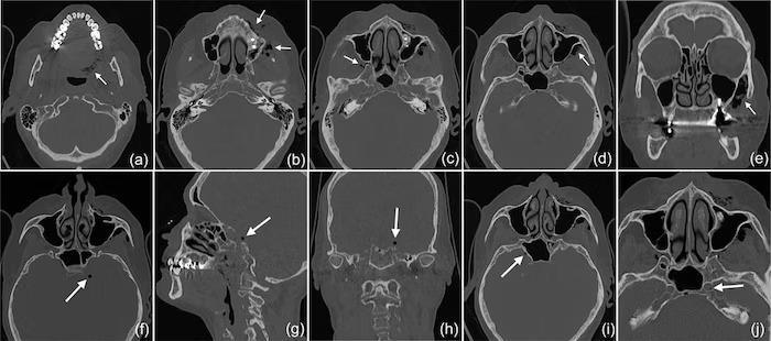
Computed tomography showed that subcutaneous emphysema had spread to the left side of the woman's pharynx (a) and face (b) into the right (c) and left (d) pterygopalatine fossae with a maximum expansion of up to 3 cm on the left side (e). Intracranial air bubbles were up to 5 mm in diameter (axial [f], sagittal [g], and coronal [h] views). Bubbles were found on both sides of the sella turcica in the region of the cavernous sinus (i) and on the left side in the carotid canal (j).
Due to the risk of developing meningitis, the woman was immediately admitted to the hospital. She was closely monitored and received intravenous antibiotics three times a day. CT showed that the pneumocephalus had resolved and the soft tissue emphysema had regressed. She was discharged three days after admission with a prescription for oral antibiotics for 5 days. Her recovery was unremarkable.
A few months after her hospital stay, the pocket depth on Implant 27 deteriorated and she decided to have it removed. The operation and postoperative recovery were uneventful.
Identification of a possible cause
Since 1960, approximately 150 cases have been reported in which patients developed subcutaneous emphysema associated with dental procedures. Emphysema was found in various places, including the mediastinum and orbital region. In many cases, emphysema has occurred after the use of high speed handpieces and air syringes.
This is believed to be the first case of pneumocephalus developing during mucosal treatment of a peri-implant lesion with an air polisher. The authors suspect that bone defects may have played a role in this particular case.
CT showed a deep peri-implant bone defect on the mesial side of Implant 27 that extended almost into the sinus space, as well as a bone defect on the sinus wall that was likely a remnant of a previous sinus floor elevation procedure.
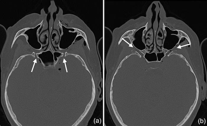
CT shows that pterygoid canal (a) and foramen magnum (b) may be ways of getting air into the intracranial space. It is especially likely in the channel leading to the round hole, where several smaller air bubbles were trapped.
“Consequently, because the nozzle was inserted on the mesial side of the implant, compressed air could spread along the peri-implant defect and then exit through this residual bone defect in the lateral wall of the sinus and into the surrounding soft tissues of the facial side of the posterior left maxilla,” the authors wrote. edura-sprovocirovala-pnevmocefaliju-u-zhenshhiny-2fbce40.jpg” alt=”A dental procedure provoked pneumocephalus in a woman3″ />
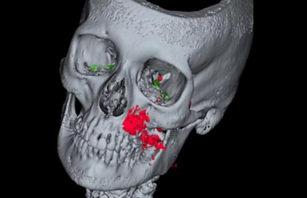
3D reconstruction of emphysema (red) and mapping of potential pathways to the intracranial space, including along the inferior orbital fissure and through the foramen magnum (green) and/or the pterygoid canal (yellow).
More and more patients decide to install implants. Accordingly, peri-implant complications are becoming more frequent, as is the use of air polishing devices in the treatment of these conditions. Therefore, clinicians should expect emphysema to occur more frequently.
More research is needed to provide evidence-based recommendations for the use of the air polishing handpiece in the treatment of peri-implantitis. In addition, if subcutaneous emphysema is identified, more extensive imaging should be considered so that the risk of complications can be assessed.
“It is suggested that in the case of extensive subcutaneous emphysema, a more extensive x-ray examination, including the craniocerebral and mediastinal space, should be considered as a standard examination in order to assess the risk of potential life-threatening complications,” Bruckmann and her colleagues concluded.