
A 51-year-old man accidentally swallowed a dental crown, leading to an unusual case of appendicitis caused by swallowing a foreign body.
Imaging, in particular X-ray and Cone Beam Computed Tomography (CBCT), has played a vital role in identifying a foreign body and assisting clinicians in treatment planning. The man has already fully recovered after the removal of the dental crown, but the ingestion of a foreign object that caused appendicitis makes this case rare and important.
“More specifically, what is unique about this case of appendicitis is that inflammation of the appendix of the caecum (appendix) is caused by a dental crown that the patient mistakenly swallowed,” write the authors, led by Zachary Brennan, a medical student in the Department of Surgery at the College of Osteopathic Medicine. Medicine at Michigan State University at East Lansing.
Typical manifestation, rare cause
A 51-year-old man presented to the emergency department after experiencing severe pain that was localized in the right lower quadrant of the abdomen for several days. According to the report, he reported experiencing nausea, vomiting, and a sharp, increasing pain in his abdomen.
The man's medical and surgical history was unremarkable, but he recently visited a dentist to have crowns placed on six of his teeth. The patient reported that most of his old crowns had fallen out. He admitted that he may have swallowed one of them.
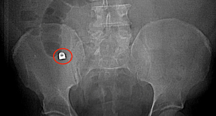
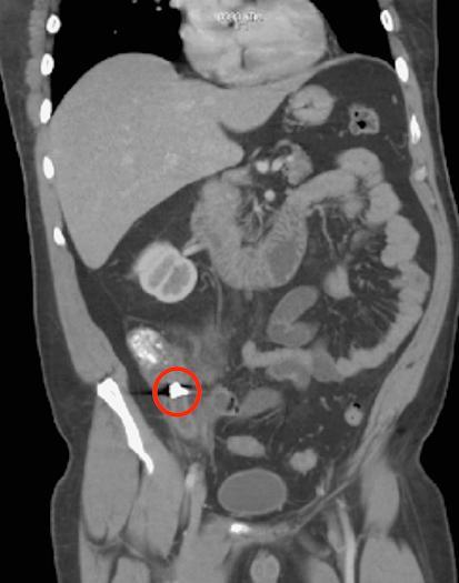
On physical examination, the man had no fever, normal pulse, and normal blood pressure. But the number of leukocytes in the blood was increased. Due to suspected appendicitis, emergency doctors ordered an x-ray of the abdominal cavity and computed tomography.
Imaging revealed a 1 x 0.9 cm induration in the man's appendix, as well as signs of inflammation consistent with acute appendicitis. Imaging also showed the absence of normal muscle contractions in the intestines in the man's regional right lower quadrant.
Computed tomography also confirmed the presence of a metal object – a dental crown – in the patient's appendix.
Based on the imaging results, doctors diagnosed the man with acute appendicitis. He was prescribed prophylactic preoperative antibiotics and then successfully underwent a robotic-assisted appendectomy.
After the procedure, the doctors examined the endoscopic bag and found a dental crown, which caused the man's appendicitis. According to the authors, the man recovered without complications.
Foreign object appendicitis
Foreign object appendicitis is rare, accounting for about 0.005% of total cases in the United States. After analyzing 100 years of studies of cases of appendicitis caused by foreign body ingestion, the authors noted that cases with swallowed objects cause tears, and almost all objects are radiopaque. This highlights the importance of using visualization.
When the clinical picture suggests uncomplicated appendicitis, as in this case, a detailed medical history, physical examination, and especially imaging are vital to treatment planning.
“While imaging is often not required in the initial diagnosis of acute appendicitis, it can also be vital in identifying a foreign body in the appendix, as in this patient,” Brennan and colleagues concluded.